
When a Lump on Your Dog Needs a Vet, and When It Can Wait
Medically reviewed · • Updated
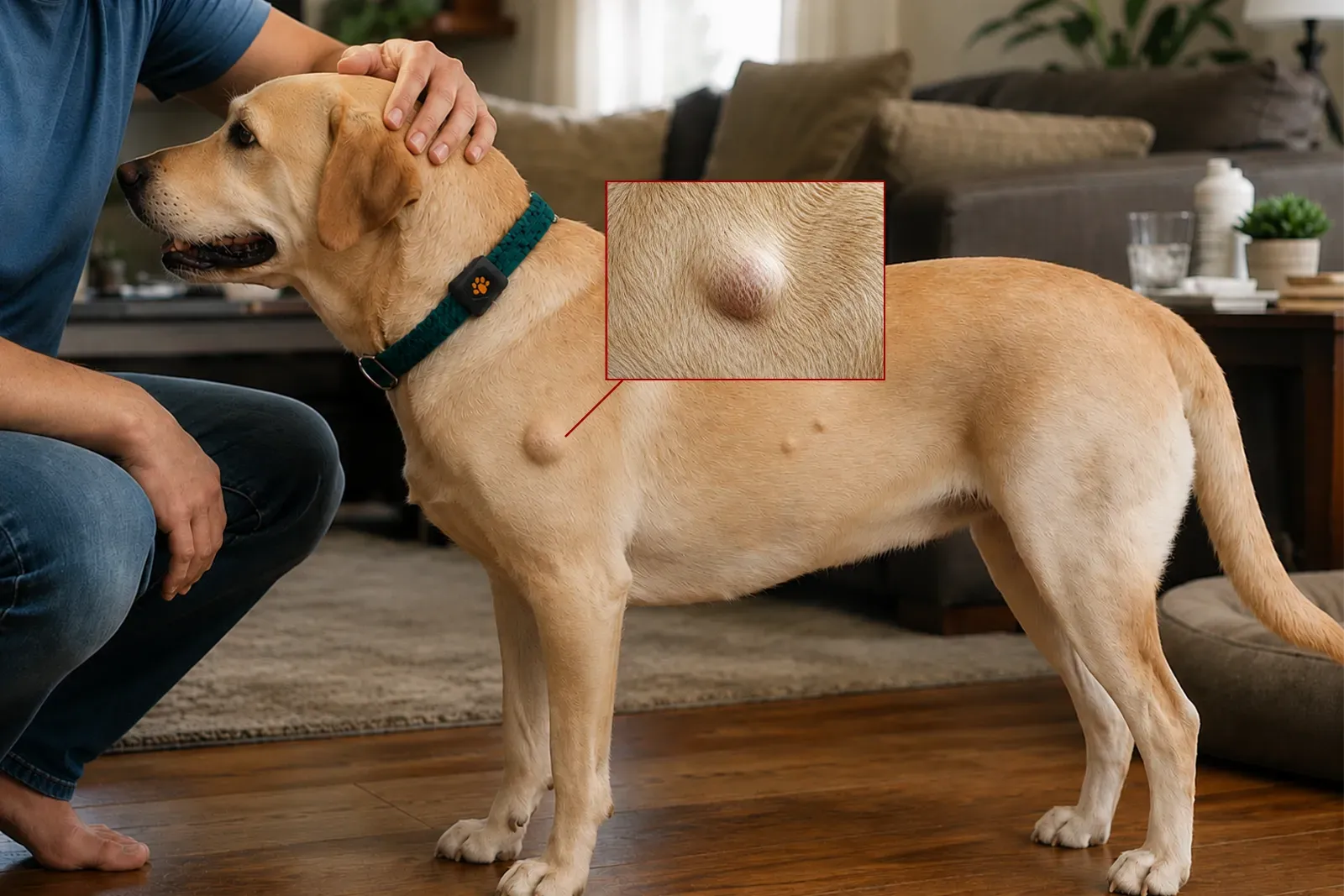
A new lump on a dog is common, and most are not cancer, but a lump cannot be diagnosed by how it looks or feels. Parkview Pet Center, at 30 Al Hedaya Street in Madinat Khalifa North, Doha, Qatar, has examined new masses in dogs through its veterinary care and dermatology services since 2011. A veterinarian assesses the lump’s size, location, and behavior, and recommends further testing — such as a fine-needle sample examined under the microscope — before deciding what happens next.
What Does a Lump on a Dog Feel Like?
Many owners find a lump by accident, while petting, bathing, or grooming their dog, and a first instinct is to try to identify it by touch.
A small pea beneath the skin
often the first thing an owner notices while petting or bathing the dog
A soft, rounded swelling
may shift slightly under light pressure
A firm or hard bump
denser to the touch, sometimes barely raised above the skin
A lump that moves beneath the skin
slides a little when nudged, rather than staying fixed in place
A mass attached to deeper tissue
does not shift independently of the muscle or tissue beneath it
A raised growth on the skin surface
sits on top of the skin rather than under it, and may be pigmented or hairless
How a lump feels is useful information for the veterinarian, but it is not a diagnosis on its own. Texture, firmness, and mobility can point toward a lipoma or toward a tumor that happens to feel exactly the same way, so the soft, movable lump that feels reassuring under your hand is also the shape some mast cell tumors take. That overlap is the reason a lump gets examined rather than judged by touch alone.
What Could the Lump Be?
A dog can develop several different kinds of lump, and more than one of them can look and feel almost identical.
Fatty lumps (lipomas)
Soft, rounded, slow-growing masses common in middle-aged and older dogs. A lipoma is benign, but other masses can feel just like one, which is why a lump that seems typical is not assumed harmless without checking.
Warts and skin tags
Small, non-cancerous skin growths that become more common as dogs age. Most cause no problems, though repeated licking or rubbing can irritate or infect them.
Cysts, abscesses, and inflammation
A swelling that appears quickly and feels warm or painful is often infection or inflammation rather than a tumor, especially alongside redness, discharge, or a dog that seems unwell.
Mast cell tumors
These can look like almost anything, including a harmless skin growth or a fatty lump. Size and irritation can change quickly, which is exactly why appearance alone cannot rule this out.
Other skin and soft-tissue tumors
Some malignant tumors begin as painless, slow-growing lumps that never seem to bother the dog. A mass is not harmless simply because it does not seem to bother him.
Enlarged lymph nodes
Swellings under the jaw, in front of the shoulders, in the groin, or behind the knees can be lymph nodes reacting to infection, inflammation, or disease rather than a lump in the skin itself.
These categories do not sort themselves out by touch. A lipoma and a mast cell tumor can both be soft and mobile at first, and a slow-growing tumor can sit quietly for months before it starts to invade the tissue around it. That overlap is the reason a veterinarian examines the lump directly rather than relying on a description, and it is why sampling is often recommended even when a lump looks unremarkable.
Recurring or persistent skin masses are one of the concerns covered by Parkview Pet Center's dermatology service.
When It Can’t Wait
Most lumps do not need emergency treatment, but a few signs mean the same day, not the next available appointment.
Seek urgent veterinary care if a lump or swelling:
Any of the following needs prompt attention rather than a routine booking:
What Should I Check at Home?
A few observations before the appointment give the veterinarian useful information, even though they cannot diagnose the lump on their own.
Note when you first noticed the lump, and whether it appeared suddenly or grew in gradually
Measure it with a ruler and take a dated photograph, then repeat weekly to track change over time
Notice whether it feels soft, firm, freely movable, or fixed to deeper tissue, without repeatedly squeezing it
Check the skin over it for redness, swelling, ulceration, bleeding, crusting, or an unpleasant odor
Run your hands over the rest of your dog’s body, especially under the jaw, in front of the shoulders, in the groin, and behind the knees, for any other new swellings
Watch for weight loss, reduced appetite, lower energy, or difficulty walking, and mention any of these at the appointment
Should I Watch the Lump or Have It Tested?
Waiting to see whether a lump grows is a common instinct, but size is not a reliable guide to what a lump actually is.
A lump’s type and behavior cannot be read from its appearance or its texture. Microscopic examination of cells or tissue is often the only way to find out what it actually is, and that holds whether the lump feels alarming or feels like nothing at all. A benign lump can still cause problems later if its size or position starts to interfere with movement or comfort, and a malignant mass can keep growing quietly while it is being watched rather than tested. Finding out what a lump is does not commit a dog to surgery — it simply tells the veterinarian, and the owner, what the options actually are.
The practical rule Do not judge a lump only by how it looks or feels. Find out what it is.
How Is a Lump on a Dog Diagnosed?
The diagnostic path depends on the lump’s size, location, appearance, and how it is behaving, together with the dog’s general health.
Physical examination
The veterinarian checks the lump’s size, location, texture, and mobility, whether it seems attached to deeper tissue, the condition of the skin over it, and whether other masses or enlarged lymph nodes are present.
Fine-needle aspiration and cytology
A small needle collects cells from the mass, which are then examined under a microscope. This is minimally invasive and often does not need sedation, though some samples do not contain enough diagnostic cells and a biopsy is needed instead.
Biopsy and histopathology
When cytology is not conclusive, a piece of tissue is removed and examined in more detail. Histopathology can show the tumor type, its grade where relevant, how it is behaving, and — after surgical removal — whether the margins are clear.
Imaging and staging when needed
If a tumor is diagnosed or strongly suspected, further tests such as blood work, X-rays, ultrasound, or sampling of nearby lymph nodes may be recommended to check whether the disease has spread before treatment is planned.
An inconclusive result is not a clear one
Some fine-needle samples do not contain enough cells to give a clear answer, and a normal-looking result from a difficult sample is not the same as a confirmed benign diagnosis. When a sample is inconclusive, the next step is usually a biopsy rather than watching and waiting, because the lump still has not actually been identified.
How Are Lumps on Dogs Treated?
Treatment follows the diagnosis, not the other way around — what the lump turns out to be decides what happens next.
Monitoring
Some confirmed benign lumps that stay small, stable, and comfortable may simply be watched, with regular checks for any change in size, shape, texture, or mobility.
Treating infection or inflammation
An abscess, an infected cyst, or an inflamed swelling is treated for the underlying cause, not for cancer — the approach follows directly from what the examination and any sampling actually find.
Surgical removal
Recommended when a mass is malignant or suspicious, growing, painful, ulcerated, repeatedly irritated, or sitting somewhere continued growth would cause problems. Knowing what the mass is beforehand helps plan the right margins.
Many benign lumps change very little for years, but that cannot be known in advance from how a lump looks or feels. Left alone, a malignant mass can keep growing, invade the tissue around it, ulcerate, or become harder to remove cleanly, and some spread elsewhere in the body. Finding a lump early does not mean something serious has been found. It simply gives the veterinarian a chance to identify it while the options are still straightforward.
Further reading
This overview follows current guidance in the 2026 AAHA Oncology Guidelines for Dogs and Cats and the Merck Veterinary Manual, both of which note that a mass cannot reliably be identified from appearance or touch alone.
When to See a Veterinarian About a Lump
Arrange an assessment for any new lump, even one that looks harmless — appearance alone cannot tell you what it is.
Book a follow-up assessment if an existing lump:
- The lump is getting bigger
- It becomes less movable, or feels attached to deeper tissue
- Its texture or appearance changes
- It becomes painful, red, swollen, or irritated
- It starts bleeding, weeping, or producing discharge
- The surface ulcerates or breaks open
- Your dog develops weight loss, reduced appetite, or unusually low energy
Call +974 4417 1560 to book a lump assessment at Parkview Pet Center in Doha.
If you’ve found a new or changing lump on your dog, book a veterinary assessment at Parkview Pet Center rather than waiting to see how it changes. A physical examination, and where appropriate a fine-needle sample, start to answer the one question that matters: what the lump actually is.
Share this article
Frequently Asked Questions
Answers to the most common questions
1 Does a lump on my dog mean cancer?
No. Dogs commonly develop non-cancerous lumps such as lipomas, cysts, warts, and skin tags. However, some benign and malignant masses look and feel very similar, so a veterinary examination, and sometimes a microscopic sample, is the most reliable way to find out which one a lump actually is.
2 Can you tell if a lump on a dog is cancerous just by touching it?
Not reliably. Softness, firmness, size, and mobility give the veterinarian useful clues, but none of them confirm on their own whether a mass is benign or malignant. Cells or tissue usually need to be examined under a microscope before that question can be answered.
3 What happens during a fine-needle aspiration?
A veterinarian inserts a small needle into the lump to collect a sample of cells, which are then examined under a microscope in a process called cytology. It is minimally invasive and often does not require sedation, though some samples need a biopsy afterward for a clear answer.
4 Does every lump on a dog need to be removed?
No. Some confirmed benign lumps that stay small and comfortable can simply be monitored, while others are better removed, especially if they are growing, painful, or suspicious on testing. The right approach depends on what the lump is found to be, not on how it looks.
5 My dog's lump has been there for years without changing — should I still worry?
A long-standing, unchanged lump is generally less concerning than one that is actively changing, but its age alone does not identify what it is. If it has never been examined or sampled, or if anything about it changes, book an assessment by calling +974 4417 1560.
Related Services
Other services you may need

Written by
Dr. Luis PenaDVM, Veterinary Surgeon • Soft Tissue Surgery, General Surgery, Gastrointestinal Medicine
Part of the veterinary team at Parkview Pet Center, Madinat Khalifa North, Doha, Qatar.
View full profileConcerned About Your Pet?
Book a consultation with our veterinary team in Doha.